History of AFib
How atrial fibrillation was first described, what each generation of physicians could see, and where the field stands today.
Atrial fibrillation is one of the oldest documented heart conditions, and also one of the most reframed. It has been called an irregular pulse, a fibrillating chamber, an electrical disorder, and a disease of the atrial wall. Each generation of physicians named AFib from the tools they had on hand, and each new tool added a layer the one before it could not see.
What Is AFib?
AFib is a disturbance in the rhythm of the heart's upper chambers, called the atria. In a healthy heart, an electrical signal travels along the nerves on a steady cadence and the atria contract on cue. In atrial fibrillation, that signal is scrambled. The atria quiver instead of contracting, the lower chambers fire on an irregular pattern, and the pulse loses its rhythm.
Patients feel AFib in different ways. Some describe a flutter in the chest, a skipped beat, or a racing pulse. Others feel only fatigue, shortness of breath, or nothing at all. The clinical picture is the same on a tracing: a heart rhythm that has lost its order at the source.
The Earliest Descriptions
AFib as a felt experience is older than modern medicine. Texts from ancient China, often grouped under the Yellow Emperor's Inner Classic, describe a chaotic and disordered pulse and link it to long term illness. Greek and Roman physicians, including Galen in the second century, wrote at length about pulse irregularity without a way to look at the rhythm itself.
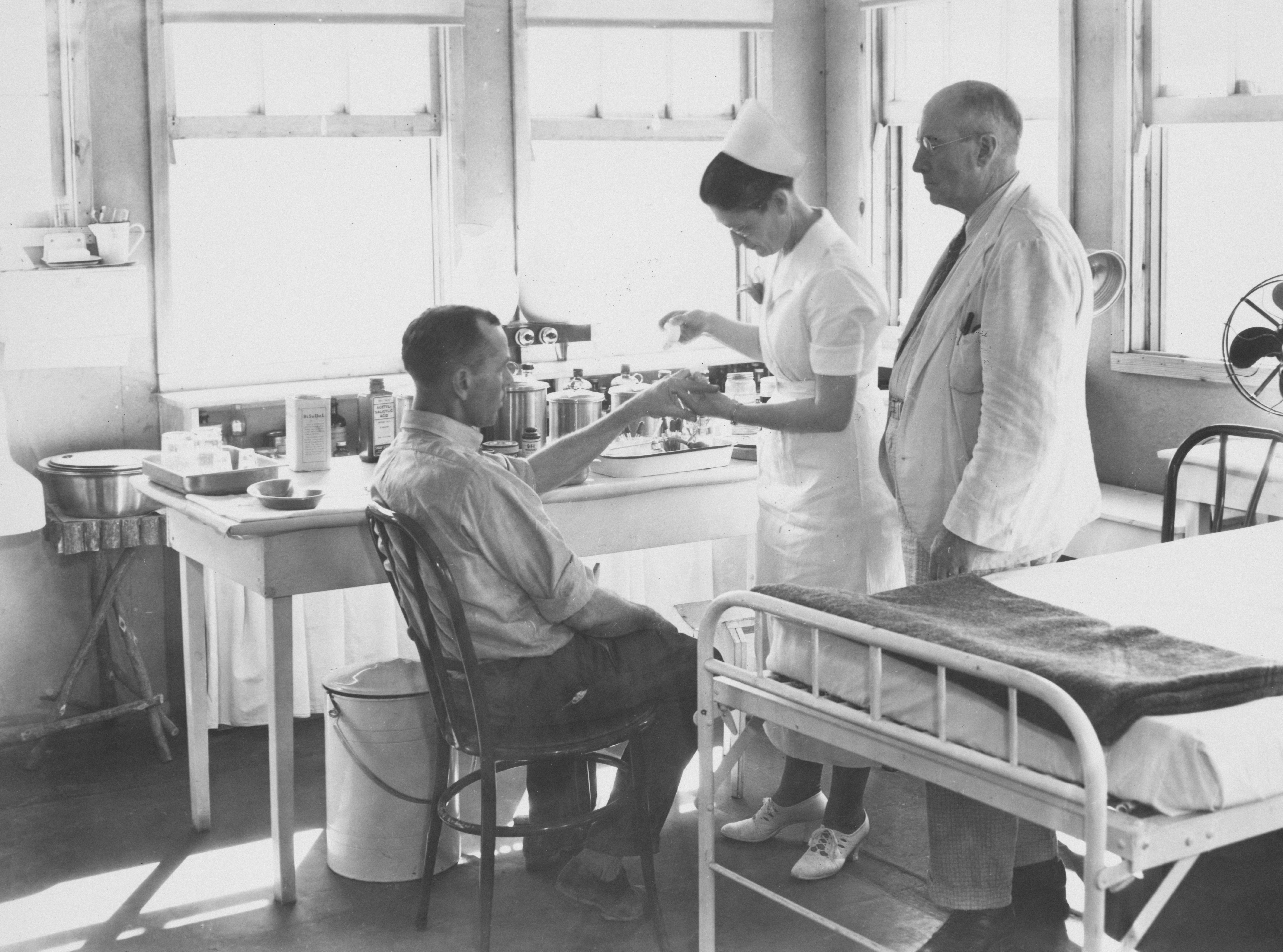
For most of the next fifteen hundred years, the pulse at the wrist was the only window into the heart. William Harvey laid out the mechanics of circulation in 1628 and Jean Baptiste de Sénac described irregular heart action in the eighteenth century, but the rhythm in the chest was still inferred rather than observed. AFib was a feeling and a finding, not yet a diagnosis.
The First Modern View
The picture changed when the heart could be recorded. In 1903, Willem Einthoven introduced the string galvanometer, the device that became the modern electrocardiogram. For the first time, the electrical activity of the heart could be drawn on paper.
In 1909, Sir Thomas Lewis published "Auricular Fibrillation: A Common Clinical Condition" in the British Medical Journal and used the new tracing to show what was happening in the upper chambers. The chaotic atrial activity that physicians had been hearing through a stethoscope finally had a fingerprint. AFib had a name, a tracing, and a place in cardiology.
What the Twentieth Century Added
Once AFib could be seen, the field began building tools to manage it. In 1918, Walter von Frey reported the use of quinidine to convert AFib back to a normal rhythm, opening the era of rhythm control medication. Digitalis, already in use for heart failure, was widely adopted to slow the ventricular rate during AFib. Warfarin and related anticoagulants moved into broad use through the 1950s and 1960s, after research linking AFib to clot formation made stroke prevention a central goal of treatment.
Direct current cardioversion arrived in 1962 when Bernard Lown described a synchronized electrical shock that could reset the rhythm in a controlled setting. The catheter era opened later. In 1998, Michel Haïssaguerre and colleagues published work in the New England Journal of Medicine showing that the muscle sleeves around the pulmonary veins were the source of many AFib triggers. That paper became the foundation for modern catheter ablation, the procedure that uses heat or cold to scar the tissue around those veins.
By the end of the twentieth century, AFib had a defined toolbox: rate control, rhythm control, anticoagulation, cardioversion, and ablation. Each tool addressed a different consequence of the condition.
How Understanding Has Shifted
For most of the twentieth century, AFib was framed as a wiring problem inside the heart. The atria were seen as the source, the pulmonary veins as the trigger, and the lower chambers as the downstream symptom.
The modern view is broader. Researchers now describe AFib as a condition shaped by inflammation, autonomic nerve activity, atrial stretch and remodeling, and fibrosis in the heart wall. The autonomic nerves that travel along the heart and tell it when to beat are part of that picture. Studies in the last two decades have documented elevated inflammatory markers in AFib patients, neural remodeling in the atria, and a strong link between long term inflammation and recurrence after treatment.
That shift matters for patients. It means AFib is no longer viewed only as faulty wiring inside the heart, and the conversation has started to include what is happening on the nerves around it.
Where Standard Care Stands Today
Modern AFib care is built around the toolbox the twentieth century produced. Rate control medication slows how fast the lower chambers respond. Rhythm control medication tries to keep the heart in a normal rhythm while the patient is taking it. Anticoagulants reduce the stroke risk that AFib creates. Catheter ablation scars the trigger sites near the pulmonary veins. Surgical ablation and pacemakers are reserved for harder cases.
Each of these is organized around a symptom. They are designed to manage the rhythm, manage the rate, or manage the clot risk that AFib produces. None of them are designed to address the inflammation along the nerves that increasingly appears in the underlying picture.
A New Frontier: Kaorta
Kaorta® is the only U.S. based patented cell therapy protocol developed for atrial fibrillation and congestive heart failure. The protocol is built around a different layer of the condition: inflammation along the nerves that tell the heart when to beat. AFib and CHF share that root driver, and Kaorta® is designed to work on it directly.
A treatment cycle begins with tissue donated by the patient or a related donor and collected by a plastic surgeon. The harvest is developed into PVC (perivascular) cells through a patented process carried out inside an FDA registered laboratory, with the patent and the laboratory held by separate entities. The cells are delivered through a series of IV infusions in a clinic setting. On each treatment day the patient relaxes in a chair while the infusion is given, then goes home the same day.
The PVC cells help the body reduce the inflammation along those nerves. As that inflammation comes down, the nerves have room to recover and the signal to the heart has a chance to steady. The therapy follows rigorous protocols, is administered in a controlled setting, and complies with FDA guidelines outlined in 21 CFR 1271.10.
For a patient or family weighing AFib options, Kaorta® is not a replacement for a cardiologist's plan. It is a different layer of the same condition, focused on the cause rather than the symptom.
Sources
- Lewis, T. "Auricular Fibrillation: A Common Clinical Condition." British Medical Journal, 1909.
- The Nobel Prize in Physiology or Medicine 1924. Willem Einthoven Facts. NobelPrize.org.
- Haïssaguerre, M., et al. "Spontaneous Initiation of Atrial Fibrillation by Ectopic Beats Originating in the Pulmonary Veins." New England Journal of Medicine, 1998.
- Lown, B., Amarasingham, R., Neuman, J. "New Method for Terminating Cardiac Arrhythmias: Use of Synchronized Capacitor Discharge." JAMA, 1962.
- Hu, Y. F., Chen, Y. J., Lin, Y. J., Chen, S. A. "Inflammation and the Pathogenesis of Atrial Fibrillation." Nature Reviews Cardiology, 2015.
- Joglar, J. A., et al. "2023 ACC/AHA/ACCP/HRS Guideline for the Diagnosis and Management of Atrial Fibrillation." Circulation, 2024.
- U.S. Food and Drug Administration. "21 CFR Part 1271: Human Cells, Tissues, and Cellular and Tissue Based Products."